
What is a Varicocele?
A varicocele is dilatation (widening) and turtuosity (irregular non-straight pass) of the veins of the testis.
The testis on yourr right hand side shows normal shape and form, while the right one has varicocele, where the veins are dilated and numoerous.

In the presence of a varicocele on one side, the other side is affected as well due to the communication between both testes. Most commonly, varicocele occurs either on both sides or on the left side. It rarely occurs on the right side solely.
At the very beginning, the testis produces sperm in a concentration higher than normal and motility / morphology are normal. Gradually, sperm production decreases both in quality and quantity, leading to decrease in sperm concentration, motility, normal forms, and above all, a decrease in fast forward motility and in the capability of fertilizing the ovum. The latter does not show in a normal semen analysis. Therefore, a normal semen in the presence of infertility and a varicocele may mean that sperm fertilization capacity is abnormal. Eventually, in long standing larger grades of varicocele, the testis becomes small and soft (atrophy), with no sperm production.
Varicocele may cause infertility regardless its grade, whether small or large. Naturally, the larger the varicocele, the faster the deterioration is. What causes varicocele? This is usually a family predisposition where there is weakness of the body walls (congenital weakness of the mesenchyme) affecting blood vessels (varicocele and leg varices) or abdominal wall (hernia). In many cases, the cause is unknown, but the onset of varicocele is aided by habitual straining such as in cases of long standing constipation, long standing cough (smokers), weight lifting and prolonged standing.
Rarely, varicocele is caused by an abdominal mass. This is suspected if varicocele occurs only on the right side. What are the symptoms of varicocele? Usually there are no symptoms and varicocele is discovered when infertility occurs. Sometimes, in large grades of varicocele, there may be pain or heaviness, or there may be a whitish drop that comes out of the penis following urination or defecation.
إشترك في قناة أ.د.أسامه شعير علي اليوتيوب ليصلك أحدث المقاطع بصورة متجددة و فورية
Treatment of Varicocele
Regardless its size, all grades of varicocele (mild, moderate or sever) can cause infertility, though to different extents. Pain, however, is usually a result of the larger grades of varicocele. Treatment of varicocele is essentially by surgery (varicocelectomy), which is a one-day procedure that does not usually require hospital stay for more than a couple of hours, and does not usually require more that one day bed rest.
When it comes to large grades of varicocele (Grade 3), there is a consensus that it should be treated surgically, otherwise it will result in total dysfunction of the testis (atrophy). Mild and moderate varicocele (grade 1 and 2) may not cause atrophy of the testis but do cause infertility that increases by time as long as the varicocele is left untreated, which is why it should be surgically eliminated.
Varicocelectomy (surgical treatment of varicocele) is performed by interrupting the abnormal veins in which blood pools, leaving behind the normal veins through which blood flows normally. Interrupting the continuity of abnormal veins is performed by closing the vein at two close points by sutures, and cutting the vein in-between the two points.
The next figure shows the incision for “subinguinal varicocelectomy”, which is one of the most favored surgical approaches due to the very short convalesence. The incision is situated to one side of the penis.
The incision is mostly 2-4 cm long, operative time ranges from 30-40 minutes, and the patient may leave the hospital in the same day and may return to light work the next day or the day after, with few exceptions.
Time needed: 30 minutes
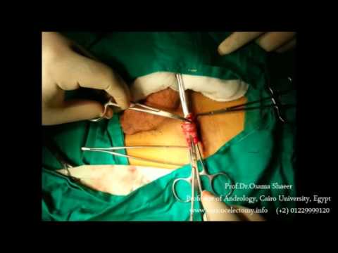
Treatment of Varicocele ; Steps of Varicocelectomy
- Incision
For subinguinal varicocelectomy, the incision is 2-4 cm long above the neck of the scrotum

- Identification
Under optical magnification and possibly using microdoppler, the different structures in the spermatic cord are identified (the veins, nerves, artery and vas deferens)

- Varicocele ligation
Surgical sutures are passed around the varicocele veins, protecting the other structures. The sutures are tied to close the dilated abnormal varicocele veins.
,
Alternative surgical and non-surgical approaches to the treatment of varicocele:
1-Open Surgery:
As shown in the illustration, one of three incisions can be used, each having its pros and cons. For example, the lowest incision is the point of meeting of all possible vein systems, while the highest incision provides access to only one system of veins (three are three systems). On the other hand, the highest incision provides access to the veins and not the arteries which keeps the arteries safe, while the lowest incision provides access to all vessels which necessitates avoiding the arteries. The two high incision involve opening the abdominal muscles, while the lowest does not, leading to easier recovery and less pain.
However, varicocelectomy should NEVER be done in the scrotum.
2-Laparoscopy:
This is endoscopy of the abdomen, that is performed through three separate incision, 1 cm each. The virtue of endoscopy is that it provides access to the right and left varicocele through the same small incisions. However, it provides access to only one system of veins, and the other two systems cannot be accessed. It is therefore that laparoscopic varicocelectomy is reserved for lower grades of varicocele when present on both sides
What are the latest advances in varicocelectomy
Time line:
Varicocele treatment changed radically with the advent of microsurgery. However, not until the introduction of intra-operative microdoppler that varicocelectomy became totally safe, artery-preserving
Microsurgical varicocelectomy
As stated in the section on “What could go wrong”, injuring the normal structures during surgery is possiple, mosst importantly the artery, due to its very small size; less than one mm.
Microsurgical varicocelectomy is the use of optical magnification to magnify the artery and veins, whereby the pulsations of the artery are more likely to be visible, so as to identify it and preserve it.
This requires special experience with microsurgery, as well as relatively costly microsurgery equipment that may not necessarily be present in every health care facility.
Intra-Operative Microdoppler for varicocelectomy

This is the latest advance in the field of varicocele treatment. In many cases, despite the use of microsurgical varicocelectomy technique, the artery may not be identified. This is because the artery is very similar to the smaller veins in shape and color, and can only be identified if pulsations are visible, which may not be the case in some instances.
Intra-Operative Microdoppler ensures that the artery is accurately identified in every varicocele surgery. Intra-Operative Microdoppler is a sterile pen-like probe that when touching a blood vessel, it sounds-out the waves of blood flow within, thereby differentiating between arteries and veins. Flow waves inside the arteries are pulse-like. Flow waves within veins are continious.
This is considered the latest and most effective development in the field of surgical treatment for varicocele, whereby the procedure’s complications possibilities are eliminated.
Contrary to microsuigery, the use of Intra-Operative Microdoppler is very easy to learn, and the equipment is much less costly, and can even be leased in some countries.